Uric Acid Crystals in Joints & Urine: Symptoms, Diagnosis, and How to Dissolve Them
Uric Acid Crystals in Joints & Urine: Symptoms, Diagnosis, and How to Dissolve Them
Quick summary (read this first)
- Uric acid crystals are the direct cause of gout.
- Crystals form when uric acid levels exceed about 6.8 mg/dL (saturation point).
- Dissolving uric acid crystals requires keeping uric acid below saturation long enough (often <6 mg/dL for gout).
- Crystals can also form in the urinary tract (uric acid stones), especially when urine is acidic.
- If you have fever + a hot swollen joint, or severe flank pain/blood in urine, seek urgent medical care.
What are uric acid crystals?
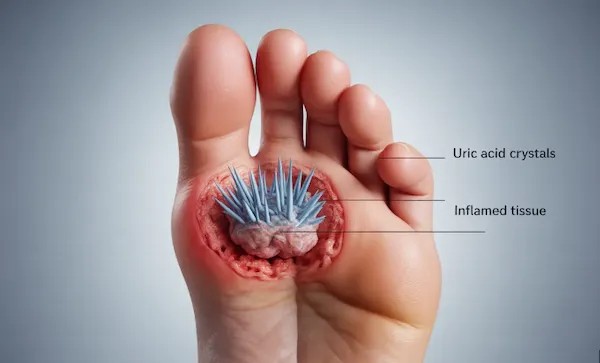
Uric acid crystals are microscopic, needle-shaped crystals that form when uric acid becomes too concentrated. In gout, the classic crystals are monosodium urate (MSU).
Crystal characteristics
- Shape: needle-like or rod-shaped
- Size: ~2–20 micrometers
- Seen under microscopy (not visible to the naked eye)
Uric acid crystals in urine vs joints
Uric acid crystals can show up in different places, and the management can differ.
In joints (gout)
- MSU crystals deposit in joints and trigger intense inflammation.
- Common sites: big toe, ankle, knee, foot joints, fingers (later stages).
- Goal: lower serum uric acid below the level where crystals form, and keep it there.
In urine (uric acid stones/crystals)
- Uric acid is less soluble in acidic urine (commonly pH <5.5).
- This can contribute to uric acid kidney stones in some people.
- Management may include hydration and (for selected patients) urine alkalinization under medical supervision.
How uric acid crystals form
The crystallization process
- Uric acid accumulation: production exceeds excretion.
- Supersaturation: levels rise above ~6.8 mg/dL.
- Nucleation: “seed” crystals form (often in cooler, lower pH, or damaged areas).
- Crystal growth: crystals enlarge as more urate attaches.
- Deposition: crystals settle in joints/soft tissue/kidneys.
Factors that accelerate crystal formation
| Factor | Why it matters |
|---|---|
| High uric acid levels | More urate available to crystallize |
| Cold temperature | Urate is less soluble in colder tissues |
| Dehydration | Concentrates urate in blood/urine |
| Acidic environment | Reduces urate solubility |
| Tissue injury | Provides nucleation sites |
| Rapid uric acid changes | Can mobilize deposits and trigger flares |
Where uric acid crystals deposit
Uric acid crystals in feet (why the big toe is common)
The big toe (podagra) is a classic first site.
Why feet are affected early
- Cooler temperature in extremities
- High mechanical stress from walking/standing
- Slower circulation compared with core body tissues
Common foot/ankle sites
- Big toe joint (MTP joint)
- Ankle
- Midfoot
- Achilles tendon area (tophi)
Joint deposits (gout)
Common joints
- Big toe (podagra)
- Ankle
- Knee
- Wrist/fingers (more common in advanced gout)
- Elbow (often with tophi)
Soft tissue deposits (tophi)
Tophi are visible or palpable deposits of urate crystals under the skin.
Common sites
- Ears
- Fingers/hands
- Elbows
- Achilles tendon
- Feet/toes
Kidney deposits
Uric acid can contribute to:
- Uric acid stones (especially acidic urine)
- Urate nephropathy (rare, typically severe settings)
Symptoms of uric acid crystal deposits
Acute gout attack (classic)
- Sudden onset, often at night
- Severe pain
- Swelling
- Redness/warmth
- Extreme tenderness (even bedsheets hurt)
Typical duration
- Untreated: ~7–14 days
- Treated: often shorter
Chronic crystal burden (between attacks)
- Stiffness
- Reduced range of motion
- Mild ongoing discomfort
- Progressive joint damage in uncontrolled disease
Kidney stone symptoms
- Severe flank/back pain
- Blood in urine
- Nausea/vomiting
- Urinary urgency/frequency
- Fever if infection occurs
Diagnosing uric acid crystals
Gold standard: joint fluid analysis
A clinician withdraws synovial fluid (arthrocentesis) and examines it under polarized microscopy.
Typical gout findings
- Needle-shaped crystals
- Negative birefringence
This can also distinguish gout from pseudogout (calcium pyrophosphate crystals).
Imaging (supportive)
- Dual-energy CT (DECT): can visualize urate deposits in some settings.
- Ultrasound: may show a “double contour sign.”
- X-ray: shows damage in later-stage gout (not early).
- MRI: detailed soft tissue imaging when needed.
Blood tests
- Serum uric acid (can be normal during a flare).
- Kidney function (creatinine/eGFR) for treatment planning.
How to dissolve uric acid crystals
Dissolving crystals requires reducing urate saturation long enough that deposits gradually shrink.
Target uric acid levels
- Many gout patients: <6 mg/dL
- Severe gout/tophi: <5 mg/dL (often used)
Timeline for dissolution (typical)
| Crystal burden | Typical timeframe (with consistent urate control) |
|---|---|
| Small deposits | 6–12 months |
| Moderate tophi | 1–2 years |
| Large tophi | 2–5 years |
| “Full clearance” | Can take 3+ years in some cases |
Medical approaches (overview)
- Xanthine oxidase inhibitors (reduce production): allopurinol, febuxostat.
- Uricosurics (increase excretion): probenecid (selected patients).
- Pegloticase (severe/refractory cases).
Medication selection depends on kidney function, drug interactions, and clinical history.
Prevent flares during dissolution
When crystals mobilize, flares can occur. Clinicians may use flare prophylaxis (e.g., colchicine/NSAIDs/steroids in selected cases) temporarily.
Removing gout crystals permanently (long-term plan)
Think in phases:
- Get below saturation (often <6 mg/dL).
- Stay there consistently (months/years).
- Reduce triggers that cause sudden urate shifts (dehydration, binge alcohol, big purine loads).
This is why “removing gout crystals” is usually not a one-week project—it’s a consistency project.
Preventing uric acid crystals
Daily prevention habits
- Hydration (steady fluids, especially in heat/exercise).
- Lower-sugar beverage pattern (avoid fructose-heavy drinks).
- Alcohol moderation (avoid beer during flare-prone periods).
- Healthy weight and steady activity.
- Long-term urate control if you have gout history.
Urine alkalinization (kidney stone prevention)
For some people with uric acid stones, clinicians may recommend strategies to raise urine pH (often potassium citrate). Do this with medical guidance.
Frequently asked questions
Q: Can you feel uric acid crystals forming?
A: Usually no. You feel symptoms when crystals trigger inflammation (a flare) or create mechanical problems (tophi/joint damage).
Q: Do uric acid crystals go away on their own?
A: Not reliably. Without sustained urate control below saturation, crystals often persist and accumu